Motion Through the First Cervical Vertebrae Is Best Described as
The cervical vertebrae are numbered in the conventional way C2-C7. The motion through the first cervical vertebrae is best described as.
Cervical Spine Neck What It Is Anatomy Disorders
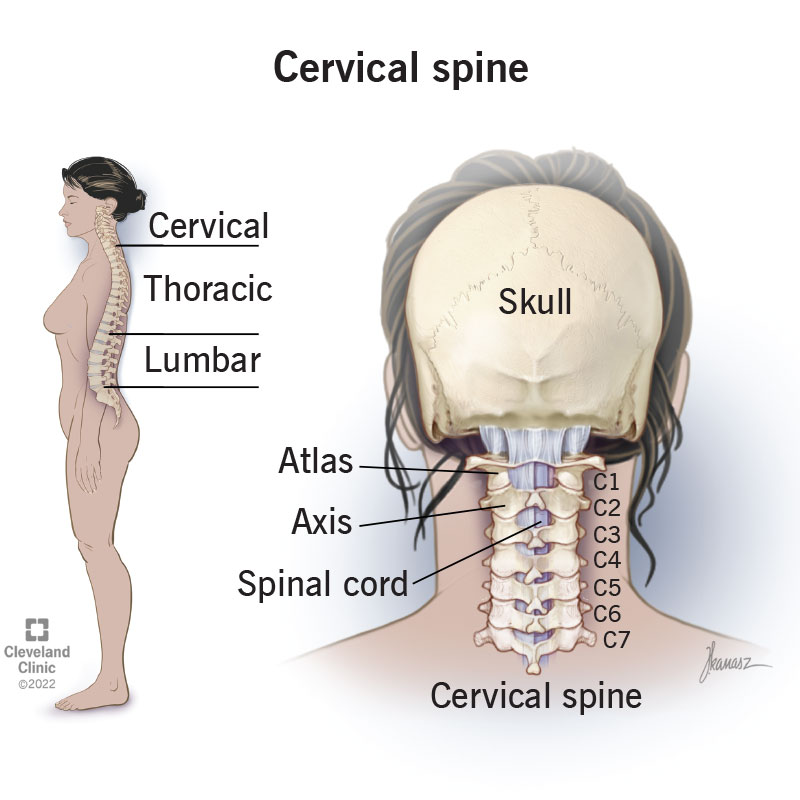
These vertebrae protect the brain stem and the spinal cord support the skull and allow for a wide range of head movement.
. The top of the cervical spine connects to the skull and the bottom connects to the upper back at about shoulder level. The cervical spine is the most superior portion of the vertebral column lying between the cranium and the thoracic vertebrae. The second cervical vertebrae C2 is known as the axis.
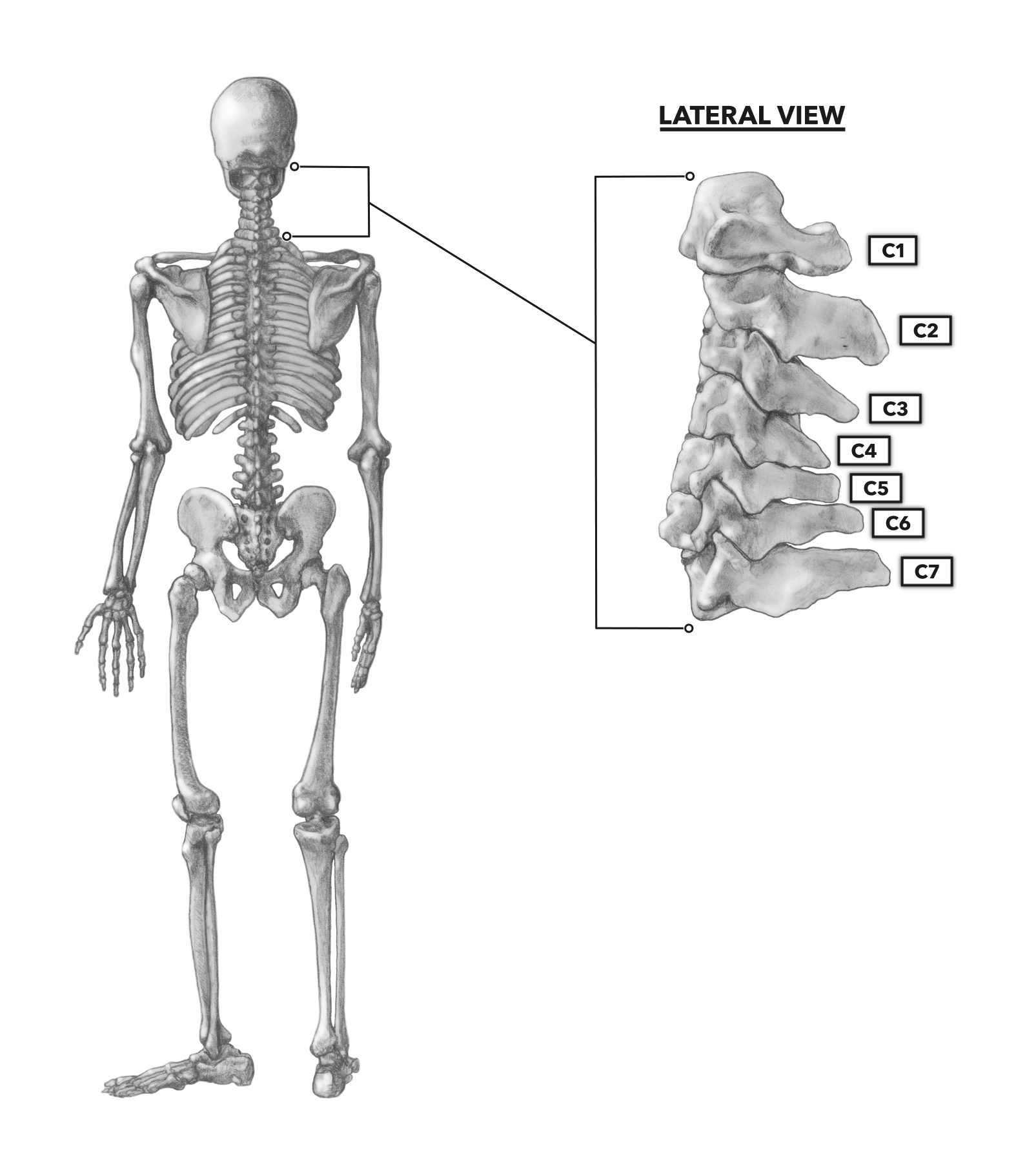
The cervical spine has 7 stacked bones called vertebrae labeled C1 through C7. The dens is a bony projection found on the first cervical vertebrae. Active motion testing by the patient through all ranges of motion.
The anterior approach to the spine first described by Robinson and Smith in their 1935 landmark paper is best employed when adequate decompression with removing a vertebral body is required. FUNCTIONAL RANGE OF MOTION. It consists of seven distinct vertebrae two of which are given unique names.
QUESTION 7 Typically how many cervical vertebrae are there located withing the vertebral column. Excessive movements of the spine by untrained and overzealous first-responders in the setting of an unstable spinal injury is one of the most common cause of a secondary CSI. The cervical spine functions to provide mobility and stability to the head while connecting it to the relatively immobile thoracic spine.
Seven cervical vertebrae labeled C1 to C7 form the cervical spine from the base of the skull down to the top of the shoulders. The first 2 C1 and C2 are highly specialized and are given unique names. According to Panjabi when a load is applied to a spinal functional unit SFU the result is a range of motion ROM which before reaching its maximum passes through a neutral zone and an elastic zoneThe neutral zone is the portion of the intervertebral mobility area closest to the rest position in which the joint has the largest capacity for movement with minimal.
The spine or vertebral column is a segmental set of 33 bones and associated soft tissues that comprise the subcranial portion of the axial skeleton. Run from transverse process of C2-C7 and attach to first two ribs will flex the spine when working together. The hardware may be placed in the front anterior or the back posterior of the Cervical spine.
1 and 2 c. This may be the main symptom. Which of the following is the best explanation for why the lateral cervical spine projection needs to be performed with a 72-inch SID.
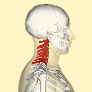
It subdivides into five regions based on curvature and morphology. At each level the cervical vertebrae protect the spinal cord and work with muscles tendons ligaments and joints to provide a combination of support structure and flexibility to the neck. The first cervical vertebrae C1 is known as the atlas.
The first vertebra in the column closest to the skull is also known as the atlas. This region consists of seven vertebrae which are abbreviated C1 through C7 top to bottom. The results can enrich current biomechanical data of cervical spine and help to find the differences.
Lab manual--exercise the axial skeleton--section the vertebral column 7 12 3 QUESTION 8 The vertebral spinal foramen is best described as. It is made up of 7 vertebrae. First the alterations in position that each osseous unit ie skull base or cervical vertebrae experienced during airway manipulation were described independently of adjacent structures yielding a value for the absolute rotational.
Atlas and axis respectively. It prevents size distortion of the cervical vertebrae 3. The first two cervical vertebrae called the atlas C1 and the axis C2 are unique even to the cervical region.
10 Spinal precautions should therefore be instituted straightaway in the pre-hospital setting to immobilize the cervical spine and minimize neck movements during the. It results in better spatial resolution of the cervical vertebrae a. The carotid-vertebral space is the surgical window between the carotid artery and the vertebral artery Fig.
The disc between the spinal bones is. The cervical spine is more mobile than the thoracic and the lumbar spine and is designed to meet the requirements of positioning of the head in space and moving to alter the visual field. The first cervical vertebra C1 is called the Atlas.
What exactly is it. Starts at C5-C6 and goes through transverse processes up through foramen magnum to supply brain. 1 and 3 d.
The rest of the cervical vertebrae C3-C7 are. Cervical Fusion is often recommended when chronic neck pain problems worsen over time. The cervical thoracic and lumbar spine the sacrum and the coccyx.
The facet apophyseal joint surfaces of most lumbar vertebrae are oriented largely in the frontal plane. It forms the joint that connects the skull and the spinal column. The neck region of the spine is known as the Cervical Spine.
Mostly flexion and extension 2nd most is sidebending little rotation. What are the symptoms of cervical pain. Cervical Fusion is a major surgery that involves joining one or more of the spinal bones together using screws bolts and plates 1.
The movement of nodding the head takes place predominantly through flexion and extension at the joint between the atlas and the occipital bone the atlanto-occipital joint. An interspace is identified by the numbers of the two surrounding vertebrae for instance C45. What is the action of the sternocleidomastoid.
Objective The aim of this study was to measure the movement of the cervical spine in healthy volunteers and patients with cervical spondylosis CS and describe the actual motion of the cervical spine using a three-dimensional 3D CT reconstruction method. 2 The transverse process of the first cervical vertebra is the most prominent of the vertebrae and is palpable deep to the superior attachment of the sternocleidomastoid muscle in patients who have a thin neck. The motion of a segment is identified by the number of the vertebra above and the motion of this vertebra is always related to the.
Cervical flexion B ipsilateral lateral flexion U and contralateral rotation U 500. The seven cervical vertebrae are the smallest and most mobile of all vertebrae reflecting the wide range of motion available to the head and neck Fig. Pain may get worse when you move your neck.
It reduces magnification of the cervical spine 2. Neck pain or stiffness. With these data cervical spinal motion during direct laryngoscopy and intubation was described in two ways.
11 Limitations in cervical ROM may restrict the ability of a person to perform those tasks that require full ROM or may. 50 There is usually minor muscle splitting in this approach and so it is considered to be more favourable for patients post-operatively. Motion at mid-cervical spine.
The cervical spine is made up of two anatomically and functionally different segments. The first thoracic vertebra is designated Thl. It is a useful.
These two segments work together to produce rotation lateral flexion flexion and extension of the head and neck. There are seven twelve and five articulating vertebrae in the cervical thoracic and.
Crossfit The Cervical Vertebrae
Cervical Vertebrae Physiopedia
Kinectmd Vertebral Column Explained The Vertebral Column Consists Of Approximately 33 Vertebra And Is Subdivided Into 5 Groups Based On Morphology And Location The Ventral Body Is The Weight Bearing

0 Response to "Motion Through the First Cervical Vertebrae Is Best Described as"
Post a Comment